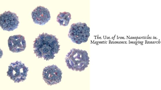
The Use of Iron Nanoparticles in Magnetic Resonance Imaging Research
Introduction
Magnetic resonance imaging (MRI) is an important medical imaging technology used to examine internal organs, tissues, and other structures without relying on ionizing radiation. As researchers continue to improve MRI performance, iron nanoparticles have attracted considerable attention for their potential role in enhancing image contrast, developing advanced imaging agents, and supporting new biomedical research applications.
Iron nanoparticles, particularly iron oxide nanoparticles, possess magnetic properties that make them useful candidates for MRI contrast agent research. Their nanoscale dimensions, tunable surface characteristics, and potential for chemical modification allow scientists to investigate new approaches to imaging, disease detection, and targeted biomedical applications.
Understanding Iron Nanoparticles in MRI Research
Iron nanoparticles are nanoscale materials containing iron, often in the form of iron oxides such as magnetite (Fe₃O₄) and maghemite (γ-Fe₂O₃). These materials can exhibit superparamagnetic behavior when their particle size and structure fall within appropriate ranges.
In MRI research, magnetic nanoparticles interact with the magnetic environment surrounding water molecules. This interaction can influence the relaxation behavior of nearby hydrogen nuclei, which contributes to the signals detected during imaging.
Depending on their composition, size, surface coating, and magnetic characteristics, iron oxide nanoparticles can affect transverse relaxation (T2 and T2*) and, in some formulations, longitudinal relaxation (T1). These effects can alter image contrast and help researchers investigate the performance of potential MRI contrast agents.
Enhancing MRI Contrast and Image Quality
One of the primary research applications of iron nanoparticles is the development of materials that improve contrast between different tissues or biological structures. Conventional MRI produces images based on differences in the magnetic properties and relaxation behavior of tissues. Contrast agents can enhance these differences, making certain structures easier to distinguish.
Superparamagnetic iron oxide nanoparticles are commonly investigated as T2 or T2* contrast agents because they can produce local magnetic field disturbances that accelerate transverse relaxation. This often results in darker regions on T2-weighted images.
Researchers study particle size, magnetic moment, concentration, and surface chemistry to understand how these properties influence contrast performance. By optimizing these variables, scientists aim to develop imaging materials that provide useful signal changes while maintaining suitable stability and biological compatibility.
The effectiveness of an iron nanoparticle formulation depends on the imaging sequence, particle characteristics, surrounding environment, and intended application. Therefore, careful experimental evaluation is essential when comparing candidate materials.
Applications in Biomedical Imaging Research
Iron nanoparticles support several areas of MRI-related investigation. Researchers explore their potential for visualizing biological processes, studying disease mechanisms, and monitoring experimental treatments.
In preclinical research, appropriately designed iron oxide nanoparticles may help investigators track labeled cells or examine the distribution of materials within biological systems. Their magnetic properties can also be studied in models of inflammation, vascular processes, and tumor biology.
Another area of interest involves combining MRI with other diagnostic or therapeutic technologies. Researchers are investigating multifunctional nanoparticles that integrate magnetic imaging capabilities with additional features, such as fluorescent labeling or drug-delivery components. Such systems may provide complementary information about biological structures and processes.
These applications remain dependent on the specific formulation and experimental evidence. Findings from laboratory or animal studies do not automatically establish safety or effectiveness for routine clinical use.
The Importance of Particle Size and Surface Modification
Particle size is a significant consideration in iron nanoparticle research because it can influence magnetic behavior, dispersion stability, and interactions with biological environments. Smaller particles may exhibit different magnetic properties from larger particles, while particle clustering can substantially change their measured performance.
Surface modification is equally important. Iron nanoparticles may be coated with polymers, silica, dextran, or other materials to improve dispersion, reduce unwanted aggregation, and modify interactions with surrounding biological components.
Researchers also investigate surface functionalization to attach targeting molecules or other chemical groups. These modifications may influence how nanoparticles interact with cells, proteins, and tissues.
For reliable MRI research, scientists typically characterize particle size distribution, morphology, chemical composition, magnetic properties, surface chemistry, and colloidal stability. These measurements help establish relationships between material structure and imaging performance.
Biocompatibility and Safety Considerations
Although iron is an essential element in biological systems, iron-containing nanoparticles cannot automatically be considered safe. Their biological effects depend on factors such as particle size, dose, coating, purity, aggregation, and route of exposure.
Research programs may evaluate cellular toxicity, oxidative stress, iron release, biodistribution, and clearance. Investigators also consider how surface coatings and impurities influence biological responses.
The stability of nanoparticles under physiological conditions is particularly important. Changes in particle structure or surface chemistry may affect their magnetic properties and interactions with biological tissues.
Before any proposed MRI contrast agent can be used clinically, it requires appropriate preclinical testing and, where applicable, clinical evaluation and regulatory authorization. Material suitability for laboratory research should not be confused with approval for human use.
Future Directions for Iron Nanoparticles in MRI
Ongoing research is exploring more precisely controlled iron nanoparticle formulations, improved surface functionalization, and methods for producing reproducible materials. Advances in nanoparticle synthesis and characterization may help researchers better understand the relationship between magnetic properties and imaging outcomes.
Multifunctional nanomaterials are another promising research direction. By combining magnetic behavior with other measurable properties, scientists may be able to investigate more comprehensive approaches to biomedical imaging and experimental diagnostics.
Further progress will depend on reproducible manufacturing, reliable characterization, appropriate biological testing, and evidence demonstrating meaningful advantages over existing imaging approaches.
Conclusion
Iron nanoparticles, particularly iron oxide nanoparticles, are valuable materials for magnetic resonance imaging research because of their distinctive magnetic properties and potential to influence MRI contrast. Their applications include contrast agent development, cellular tracking studies, experimental disease imaging, and multifunctional nanomedicine research.
Particle size, composition, surface chemistry, dispersion stability, and biocompatibility all influence their performance. Understanding these characteristics enables researchers to select and evaluate materials for specific experimental objectives.
For laboratories and research organizations investigating advanced nanomaterials, well-characterized iron nanoparticles can provide a useful foundation for exploring new MRI technologies and biomedical imaging strategies. Visit ssnano.com to explore nanomaterial options for scientific research and material development.